


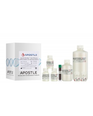
Apostle technologies have been applied in many world-class R&D studies, clinical laboratory settings, and public health response and surveillance.
This page lists some of the examples in Oncology.
For a complete list of applications citing Apostle technologies, including publications and customer testimonials, see References.
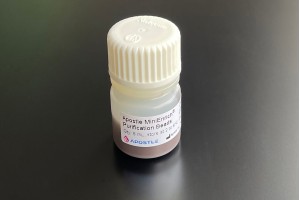
Apostle MiniMax Technology in Clinical Research on Small Cell Lung Cancer


Apostle MiniMax cfRNA Technology in Early Cancer Detection


Apostle MiniMax Technology in Clinical Research on Cancer Diagnosis

Apostle MiniMax Technology in Clinical Research on Metastatic Solid Tumors

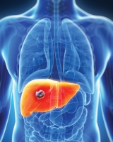
Apostle MiniMax Technology in Hepatocellular Carcinoma Detection
Apostle MiniMax Technology in Colon Cancer Detection and Post-Surgical Monitoring


Apostle MiniMax Technology in Detecting Chromosomal Structural Abnormalities in Patients with Myeloid Neoplasms


Apostle MiniMax Technology in Clinical Research of Lung Cancer Using Cell-free Chromatin Immunoprecipitation (ChIP)

Apostle MiniMax Technology in Rectal Cancer Response Prediction and Risk Stratification

Apostle MiniMax Technology in Clinical Research of NSCLC (Non-Small-Cell Lung Cancer) with dPCR
Apostle MiniMax Technology in Early Diagnosis of Lung Cancer
Apostle MiniGenomics Technology in Stool DNA Isolation and Colorectal Cancer Testing


Apostle MiniGenomics Technology in Protocols for Colorectal Cancer Screening via Analysis of DNA Methylation Biomarkers
Apostle MiniMax Technology in Nicotine Dose-Dependent Epigenomic-Wide DNA Methylation Changes in the Mice

Apostle MiniMax Technology in Circulating Cell Free RNA Analysis